Cryo-EM

— The world's leading commercial Cryo-EM center.

  • 01
    One-Stop Solution
  • 02
    Cutting-Edge Instruments
  • 03
    Experienced Teams

Characterization Of Drug Delivery Systems (LNPs, AAVs, Exosomes, etc.)

Traditional structural and morphological characterization methods for nanoparticle drug delivery carriers mainly include indirect techniques such as Dynamic Light Scattering (DLS), which typically provide information on particle size and distribution, yielding the hydrodynamic diameter. Although conventional negative-staining electron microscopy allows for direct observation of the morphology, size, and structural features of nanoparticle carriers, the results can be skewed due to staining and vacuum dehydration, which may introduce discrepancies with the sample's solution state. In contrast, cryo-EM avoids staining and enables direct observation of the sample in its native state, providing insights into both the morphology and internal structure of the sample, particularly information such as shell encapsulation rates and the localization of mRNA within lipid nanoparticle (LNP) complexes. This approach offers distinct advantages for characterizing these systems.

Advantages

  • Low sample volume requirement (30 μL,1 mg/mL concentration)

  • No sample pre-treatment required

  • Fast Turnaround (results available as soon as the same day)

  • Real morphological information obtained (especially regarding internal structure)
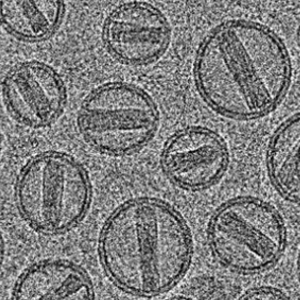
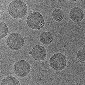
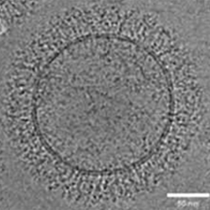
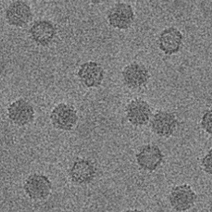
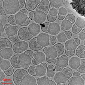
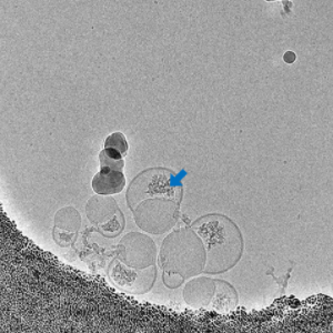
Morphology

  • API-liposome

  • mRNA-LNP

  • Exosome

  • AAV

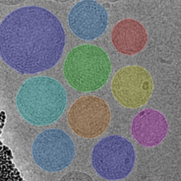
Cryo-EM Morphological Characterization of Drug Delivery Systems

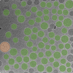
Particle Extraction and Size Distribution Statistics Based on Deep Learning

Thermo Science Amira is used to perform deep learning-based image segmentation on the cryo-EM images collected. After segmentation, the area of each particle is calculated by determining the number of pixels it occupies, which is then converted into the equivalent diameter, yielding the size distribution statistics. Additionally, the Length-to-Width Ratio (LWR) and surface rugosity of the particles can be calculated. For this type of analysis, we typically collect approximately 30 cryo-EM images and analyze a sample size of around 2,000 particles.

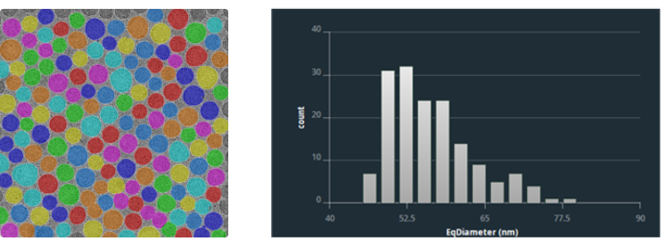
Size Distribution Statistics of Drug Delivery Systems

Membrane Thickness Analysis

For lipid-based drug-loaded liposome samples, membrane thickness is an important statistical parameter and has significant research value in understanding the morphological changes of the membrane after drug loading. After particle recognition, we outline the particle contours, then contract the contours inward. The average grayscale of each membrane layer is calculated, and the distance between the two extreme points of the average grayscale is taken as the measured membrane thickness.

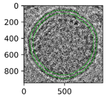
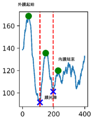
Membrane Thickness Analysis of Drug Delivery Systems

Empty-to-Full Ratio Statistics

For lipid-based samples, after image segmentation, we can calculate the average grayscale value of the pixels inside each particle. Based on this value, the particles can be classified into hollow and solid categories, allowing us to determine the ratio of hollow to solid particles.

Empty-to-Full Ratio for Lipid-based Samples

For viral vectors, due to the high uniformity of viral particles, we can collect a large number of electron microscopy (EM) images and perform large-scale analysis using single-particle analysis methods. Typically, this process involves hundreds of EM images and hundreds of thousands of particle samples. Through automated data processing, we can classify the particles into two-dimensional or three-dimensional categories to obtain the average images and the quantitative ratios of hollow and solid particles.

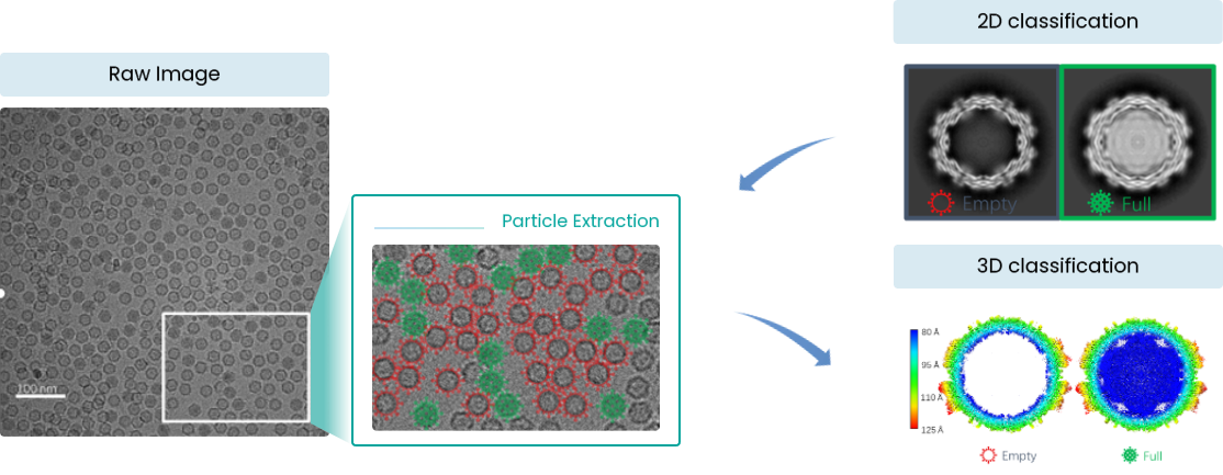
Empty-to-Full Ratio for AAV Samples

mRNA Localization by Thionine Staining

Amorphous mRNA is relatively loose and exhibits poor contrast in EM images, making it difficult to identify. Additional staining procedures are required for its localization. The staining mechanism of thioine is based on the interaction between its active functional groups and nucleic acids in the cell nucleus. The aromatic and heterocyclic structures within thioine molecule allow it to form π-π stacking interactions with the base pairs in DNA and RNA, thereby binding to the nucleic acid molecules. Additionally, the chlorine atoms and amine groups in thioine can interact with the negative charges present in the nucleic acid molecules, enhancing the binding affinity between the dye and the nucleic acids. At the EM level, the effect of thioine increases the density of mRNA, thereby enhancing its contrast in electron microscopy.
  • Before Staining

  • After Staining

  • Before Staining

  • After Staining

Localization of mRNA in LNP Samples by Thionine Staining

3D Reconstruction by Cryo Electron Tomography (cryo-ET)

Conventional cryo-EM images only provide two-dimensional projections of sample particles. For multi-layered vesicular liposomes with complex internal structures, the information obtained about their internal architecture is relatively limited. To more comprehensively analyze their internal features, we can employ cryo-electron tomography (cryo-ET) reconstruction. This technique allows for the 3D reconstruction and analysis of samples, providing more details about the internal structure.

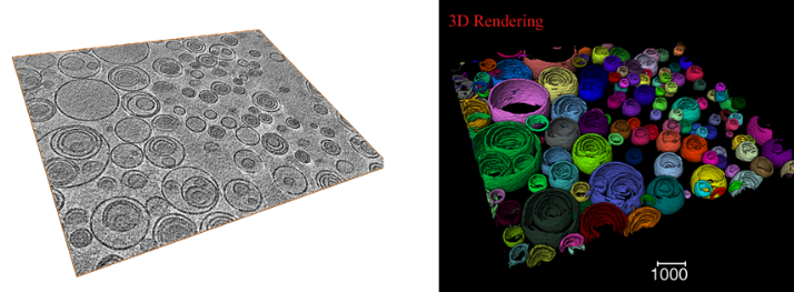
Reconstruction Results of Multivesicular Liposomes by cryo-ET

Q&A

Q

What is the typical turnaround time for sample analysis?

A

The sample preparation will be conducted on the day when sample is received, and observation results can be obtained on the same day when the sample is scheduled for the cryo-electron microscope. Statistical analysis results will require an additional 2-3 days. If the machine time is reserved in advance before submitting the sample, the waiting time can be reduced.

Q

What are the requirements for the LNP samples?

A

The total lipid concentration should generally be greater than 1 mg/mL, and the sucrose content should be as low as possible, typically not exceeding 10%.

Q

Can freeze-dried powder samples be observed?

A

Yes,but freeze-dried powder samples often have high sucrose content or low concentration after reconstitution. Typically, the samples need to be dialyzed or concentrated after reconstitution.

Q

What are the advantages of using cryo-electron microscopy to characterize drug delivery carriers?

A

Cryo-EM provides direct imaging information of the sample, especially internal structural details. The cryo-preparation preserves the native morphology of the sample, allowing for accurate particle size distribution.

Q

Are the results from DLS and cryo-EM consistent?

A

Since DLS measures the hydrodynamic diameter in liquid, while cryo-EM provides the actual particle size, the DLS results will be slightly larger. However,the overall distribution trends are consistent.

Q

I heard that cryo-EM can achieve a resolution of 3Å.Can mRNA molecules inside LNP samples be directly observed in EM images?

A

The resolution we refer to usually pertains to single-particle analysis, which requires orientation determination and averaging of hundreds of thousands of particles. This method assumes that the particles are homogeneous. However, the mRNA within LNPs does not exhibit such homogeneity, as each mRNA molecule may have a different structure. Therefore, it is not possible to perform particle alignment and averaging for these molecules.