Structural Biology
Cryo-EM (Cryo-electron microscopy)

Cryo-EM Expertise at Biortus
The Noble Prize in Chemistry awarded to Cryo-EM in 2017 has given significant recognition to the imaging power of this technique for biomolecules. Attaining atomic resolution biomolecular structures using cryo-EM is now a ubiquitous reality. Biortus sees great potential in cryo-EM for its use in structure-based drug discovery. From large protein/protein complexes, membrane proteins, proteins with dynamic conformation to viral structures and epitope mapping, structures of these targets that are “unfriendly” toward X-ray crystallography could have been solved by cryo-EM.
A wealth of Cryo-EM expertise is at hand in Biortus. Our cryo-EM team has a combined experience of over 35 years and published many cryo-EM related research in high impact journals. They are the people to talk to for an alternative to X-ray structures. Syncretizing our cryo-EM expertise with access to high-end cryo-EM equipment, Biortus is aspired to becoming the pioneering CROs in providing high resolution cryo-EM structures for clients.
What is single-particle Cryo-EM?
Cryo-electron microscopy (cryo-EM) is an increasingly popular method used by structural biologists to resolve structure of a biomacromolecules (single particles), such as protein, protein complex or virus, etc., at atomic or near-atomic resolution. The particles suspended in solution are vitrified at cryogenic temperatures and photographed by transmission electron microscopy. Usually, thousands (or even a million) of 2D images of particles in micrographs are extracted and are “merged” (based on the orientation of each particle) into a high-resolution 3D structural model of the biomacromolecule in a computer where the structure information of the particle is obtained.
Why do you need Cryo-EM Services?
Now single-particle cryo-EM technique can almost routinely deliver structure determination of well-behaved protein macromolecule at near-atomic resolution. Compared with other methods of macromolecule structure determination, such as x-ray crystallography, cryo-EM holds a number of unique advantages:
The bottleneck procedure of crystallization is not needed for sample preparation, which is a biggest advantage for some non-crystallizable proteins, such as membrane proteins.
Specimen are examined in their near-native and hydrated state.
Needs only a small amount (micro grams) of material, big advantage for low-yield specimens.
Can examine macromolecules in different functional states, and can demonstrate various conformational states in one physiological condition.
Can examine structures of large protein complexes even though they have both component and conformational heterogeneity.
Why choose Biortus for CryoEM?
Biortus has an expert team of cryo-EM scientists with very strong academic and industry experience.
Biortus delivers one-stop service all the way from gene synthesis to cryo-EM structure.
Biortus has a very strong protein production and purification team, which is essential for the rapid iteration process of sample and vitrification optimization.
Our CryoEM Workflow: From Sample to Structure
· Sample preparation:
Sample preparation is the most important step in the whole workflow. We prefer the protein is produced and purified at Biortus, so that the sample is fresh and greatly enhance the success rate for high quality cryo-EM data collection. Also, the fast feedback between protein production and cryo-EM platform ensures the fast sample optimization.
Negative staining:
Negative stain was used to check whether the sample is sufficiently pure and homogeneous in size and morphology. 2D classification and 3D reconstruction were also performed to further evaluate the state of these particles.
· Sample vitrification:
Vitrification is a process to convert solution into glass-like ice and protein particles are embedded and arrested into their near-physiological states. Different pH, buffer, grids, blotting condition, and additives will be optimized to achieve optimal vitrification results.
· High-resolution data acquisition:
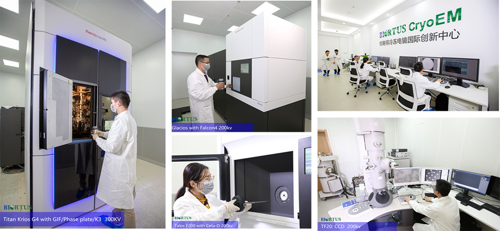
A high-resolution data collection will be performed on world-class fully-loaded Titan Krios electron microscope by highly experienced operators. Biortus can guarantee immediate and sufficient EM time without any delay.
· Data processing & 3D reconstruction:
Data processing & 3D reconstruction includes motion correction, CTF estimation, particle picking, 2D classification, 3D classification, 3D refinement and multiple polishing steps. The final 3D map delivers the structure at near-atomic resolution which can reveal side chains of amino acid or bound small chemical compound if existed. Clients usually are interested in the detailed structure of chemical-bound region, which may not have the best possible resolution due to conformational heterogeneity or flexibility. This region can then be focused classified among particles and locally refined, so that the local resultion will be enhanced to clearly show the density of bound small molecule.
· Model building:
Near-atomic resolution density map allows de novo modeling, which will reveal details of protein-protein or protein-compound interactions.


