
Structural Biology
Structural Biology
Structural Biology is a core strength at Biortus, where we employ the modern technologies for structural studies such as X-ray crystallography and Cryo-EM. We not only are able to provide protein structures for structure-based and fragment-based drug discovery, but also differentiate ourselves from other CROs by working on difficult structures like membrane proteins, large protein complexes, and epitope mapping.
Biortus constantly strives to provide timely, accurate and high-quality structural data, which made us one of the preferred partners of many global pharmaceutical companies for structure-based drug design (SBDD) services. Our crystallography team has delivered over 2000 crystal structures, and our cryoEM team has also provided more than a dozen high resolution structures to our clients.
Recently, we have developed the microED technology which allows us to further determining structures of tiny crystals of small molecules. This technology grants our reach into.
Show Cases: Epitope Mapping by X-ray Structure and cryo-EM
Mapping the actual epitope not only helps researchers to optimize their antibody drugs, but also helps them to protect patents with strong supporting data. Among many methods that determine epitopes, X-ray crystallography has always been the gold standard approach because it allows the clear visualization of the interaction between antigen and antibody. Recently, cryoEM technology has risen to be another powerful tool to obtain such information. Biortus can help clients obtain antibody epitope information determined by both X-ray crystallography and Cryo-EM technologies.
Epitope Mapping by Crystal Structure
Applications:
-
High quality epitope information (resolution better than 3.0A)
-
For patent application and substantiation
-
Antibody drug optimization
Features:
-
Mature Methodology (Gold Standard)
-
Standardized Workflow
-
High Resolution
Work Flow:
|
Experiment |
Time |
Deliverables |
|
Antigen and Antibody Preparation |
Project Dependent |
Antigen/Antibody (>90% Purity) |
|
Fab Preparation |
1- 2 Weeks |
Fab (>90% Purity) |
|
Antigen/Fab Complex Crystals |
2- 4 Weeks |
Single Crystals of Good Size |
|
X-ray Diffraction Data Collection |
2-4 Weeks |
High Resolution Structures (<3.0A) |
|
Complex Structure Determination |
1-2 Weeks |
Details of Epitope |
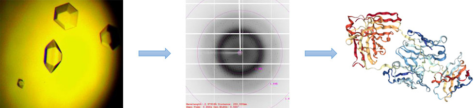
Case Study:
-
Antigen/Antibody Treatment
-
Crystal Growth/Optimization
-
Data Collection
-
Structure Solution (P21;2.83A)
-
Epitope Determined
Epitope Mapping by Cryo-EM
Applications
-
Antibody screening
-
Fast-growing alternative methodology to crystal structure
Features:
-
Faster structure delivery than crystal structure
-
Direct imaging of materials
-
Useful for very large proteins or complexes
-
Could reach 3A with recent development on both instruments and software
Work Flow:
|
Experiment |
Time |
Deliverables |
|
|
Antigen and Antibody Preparation |
Project Dependent |
Homogenous Antigen/Antibody (>90% Purity) |
|
|
Antigen/Antibody Complex Sample Preparation |
1 Day |
Vitrified Samples Ready for Electron Microscopy |
|
|
Sample Examination and Data Collection |
2-8 Weeks |
Electron Micrographs |
|
|
Data Processing and 3D Reconstruction |
4-10 Weeks |
3D Structures and Details of Epitope |
|
Case Study (Fab Fragment against a Viral Capsid Protein):
-
EM Imaging
-
3D Electron Density Map (3.8A)
-
Antibody Antigen Fitting
-
Epitope Determined
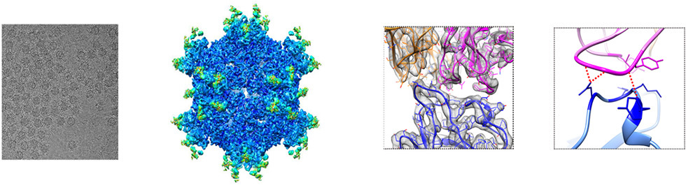
Figure Adapted from Longfa Xu et al. Nature Communications 2017. a Publication of which a Member of the Biortus cryo-EM Team (XiaoDong Yan) is a Corresponding Author
Telephone:+86 0510-86130005
Adresse:A5, 6 Dongsheng W. Rd., Jiangyin, Jiangsu, China
E-mail:info@biortus.bio
NEWS FEEDS
FOLLOW US

Copyright © 2020 Biortus Biosciences Co.,Ltd. Power by www.300.cn